
 43 Lessons
 35 Students



Oussama Lanbache
It's was perfect
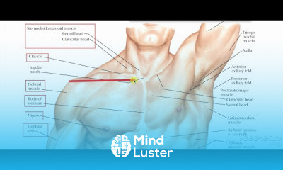
The pectoral region is the anterior region of the upper chest where there are four thoracoappendicular muscles (also known as the pectoral muscles): pectoralis major pectoralis minor subclavius serratus anterior The breast is located superficial to the muscles. The lateral border of the pectoralis major muscle forms the anterior margin of the axilla. At the superolateral aspect the pectoral region is separated from the deltoid muscle by the deltopectoral groove and contains the cephalic vein. What organs are in the pectoral region? The chest is the area of origin for many of the body's systems as it houses organs such as the heart, esophagus, trachea, lungs, and thoracic diaphragm. The circulatory system does most of its work inside the chest. The pectoral region is located on the anterior chest wall. It contains four muscles that exert a force on the upper limb: the pectoralis major, pectoralis minor, serratus anterior and subclavius. In this article, we shall look at the anatomy of the muscles of the pectoral region – their attachments, actions and innervation. Pectoralis Major The pectoralis major is the most superficial muscle in the pectoral region. It is large and fan shaped, and is composed of a sternal head and a clavicular head: Attachments: The distal attachment of both heads is into the intertubercular sulcus of the humerus. Clavicular head – originates from the anterior surface of the medial clavicle. Sternocostal head – originates from the anterior surface of the sternum, the superior six costal cartilages and the aponeurosis of the external oblique muscle. Function: Adducts and medially rotates the upper limb and draws the scapula anteroinferiorly. The clavicular head also acts individually to flex the upper limb. Innervation: Lateral and medial pectoral nerves. By TeachMeSeries Ltd (2021) Fig 1 - The sternal and clavicular heads of the pectoralis major. Fig 1 – The sternal and clavicular heads of the pectoralis major. Pectoralis Minor The pectoralis minor lies underneath its larger counterpart muscle, pectoralis major. Both muscles form part of the anterior wall of the axilla region. Attachments: Originates from the 3rd-5th ribs and inserts into the coracoid process of the scapula. Function: Stabilises the scapula by drawing it anteroinferiorly against the thoracic wall. Innervation: Medial pectoral nerve.
 To Register in Course you have to watch at least 30 Second of any lesson
 Identify the key muscles of the pectoral region and their anatomical attachments for clinical assessment and surgery
Describe the actions and innervation of pectoral muscles to understand upper limb movement and nerve injuries
Explain the relationship between the pectoral muscles, breast, and axillary structures for accurate diagnosis and surgical procedures
Analyze the anatomical features of the pectoralis major and minor to assist in muscle injury rehabilitation and physiotherapy
Recognize the neurovascular structures in the pectoral region, including the cephalic vein and pectoral nerves, for safe injections and surgeries
Illustrate the anatomical boundaries of the pectoral region to aid in clinical examinations and regional anesthesia techniques
Assess the role of pectoral muscles in upper limb movements such as adduction, medial rotation, and flexion for sports and physical therapy
Differentiate between superficial and deep muscles of the pectoral region to improve surgical planning and imaging interpretation
Apply knowledge of pectoral anatomy to diagnose thoracic and shoulder injuries accurately in clinical practice
Evaluate the relationship between the pectoral muscles and thoracic organs like the heart and lungs for thoracic surgeries and emergencies
Describe the significance of the deltopectoral groove and cephalic vein in venipuncture and surgical approaches
Utilize anatomical knowledge of the pectoral region to enhance understanding of regional anatomy in medical education and training
43 Lessons • 3 h 30 m total

 You must have an
Account
on the platform.
Complete all lessons.
Watch at least 50% of Lesson Duration.
Get your Certificate free 🎉 !
4.3
 (41 reviews)
Overall rating

Be the First One Review This Course
It's was perfect
easy to understand

really nice and complete
It will be better to have an exam after the lecture

The pectoral region is the anterior region of the upper chest where there are four thoracoappendicular muscles (also known as the pectoral muscles): pectoralis major pectoralis minor subclavius serratus anterior The breast is located superficial to the muscles. The lateral border of the pectoralis major muscle forms the anterior margin of the axilla. At the superolateral aspect the pectoral region is separated from the deltoid muscle by the deltopectoral groove and contains the cephalic vein. What organs are in the pectoral region? The chest is the area of origin for many of the body's systems as it houses organs such as the heart, esophagus, trachea, lungs, and thoracic diaphragm. The circulatory system does most of its work inside the chest. The pectoral region is located on the anterior chest wall. It contains four muscles that exert a force on the upper limb: the pectoralis major, pectoralis minor, serratus anterior and subclavius. In this article, we shall look at the anatomy of the muscles of the pectoral region – their attachments, actions and innervation. Pectoralis Major The pectoralis major is the most superficial muscle in the pectoral region. It is large and fan shaped, and is composed of a sternal head and a clavicular head: Attachments: The distal attachment of both heads is into the intertubercular sulcus of the humerus. Clavicular head – originates from the anterior surface of the medial clavicle. Sternocostal head – originates from the anterior surface of the sternum, the superior six costal cartilages and the aponeurosis of the external oblique muscle. Function: Adducts and medially rotates the upper limb and draws the scapula anteroinferiorly. The clavicular head also acts individually to flex the upper limb. Innervation: Lateral and medial pectoral nerves. By TeachMeSeries Ltd (2021) Fig 1 - The sternal and clavicular heads of the pectoralis major. Fig 1 – The sternal and clavicular heads of the pectoralis major. Pectoralis Minor The pectoralis minor lies underneath its larger counterpart muscle, pectoralis major. Both muscles form part of the anterior wall of the axilla region. Attachments: Originates from the 3rd-5th ribs and inserts into the coracoid process of the scapula. Function: Stabilises the scapula by drawing it anteroinferiorly against the thoracic wall. Innervation: Medial pectoral nerve.
Experience the full power of Mindluster with Premium.
No distractions, just pure education.
Cancel your subscription anytime