
 18 Lessons
 60 Students



Bhagwat RAM Barar
Very good program
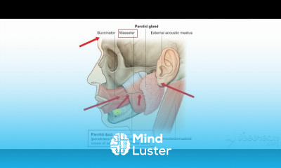
Parotid tumors are abnormal growths of cells (tumors) that form in the parotid glands. The parotid glands are two salivary glands that sit just in front of the ears on each side of the face. Salivary glands produce saliva to aid in chewing and digesting food. There are many salivary glands in the lips, cheeks, mouth and throat. Tumors can occur in any of these glands, but the parotid glands are the most common location for salivary gland tumors. Most parotid tumors are noncancerous (benign), though some tumors can become cancerous. Parotid tumors often cause swelling in the face or jaw that usually isn't painful. Other symptoms include numbness, burning or prickling sensations in the face, or a loss of facial movement. Parotid tumor treatment is usually with surgery to remove the tumor. If the tumor contains cancer cells, additional treatments might be recommended. The parotid gland is a major salivary gland in many animals. In humans, the two parotid glands are present on either side of the mouth and in front of both ears. They are the largest of the salivary glands. Each parotid is wrapped around the mandibular ramus, and secretes serous saliva through the parotid duct into the mouth, to facilitate mastication and swallowing and to begin the digestion of starches. There are also two other types of salivary glands; they are submandibular and sublingual glands Sometimes accessory parotid glands are found close to the main parotid glands. The parotid gland is a bilateral salivary gland located in the face. It produces serous saliva, a watery solution rich in enzymes. This is then secreted into the oral cavity, where it lubricates and aids in the breakdown of food. In this article, we shall look at the location, vasculature and innervation of the parotid gland. We shall also consider any clinical correlations. Anatomical Position The parotid gland is a bilateral structure, which displays a lobular and irregular morphology. Anatomically, it can be divided into deep and superficial lobes, which are separated by the facial nerve. It lies within a deep hollow, known as the parotid region. The parotid region is bounded as follows: Superiorly – Zygomatic arch. Inferiorly – Inferior border of the mandible. Anteriorly – Masseter muscle. Posteriorly – External ear and sternocleidomastoid. The secretions of the parotid gland are transported to the oral cavity by the Stensen duct. It arises from the anterior surface of the gland, traversing the masseter muscle. The duct then pierces the buccinator, moving medially. It opens out into the oral cavity near the second upper molar.
 To Register in Course you have to watch at least 30 Second of any lesson
 Identify the anatomical location and structure of the parotid gland in relation to surrounding facial features and muscles
Describe the vascular supply and innervation pathways of the parotid gland for clinical relevance and surgical procedures
Explain the clinical presentation and symptoms associated with parotid tumors, including benign and malignant types
Assess diagnostic techniques and imaging modalities used to evaluate parotid gland tumors and their effectiveness
Outline the surgical approaches and considerations for parotid gland tumor removal, emphasizing facial nerve preservation
Discuss potential complications and postoperative management strategies following parotid gland surgery
Evaluate the role of histopathology in differentiating between benign and malignant parotid tumors for treatment planning
Apply knowledge of parotid gland anatomy and pathology to identify clinical signs and develop appropriate treatment plans
18 Lessons • 1 h 26 m total
 You must have an
Account
on the platform.
Complete all lessons.
Watch at least 50% of Lesson Duration.
Get your Certificate free 🎉 !
4.5
 (8 reviews)
Overall rating

Be the First One Review This Course
Very good program
Very good

good

A nice way to make people aware.
excellent

How to able to watch in English
Very good
Can we study in English also?
excellent
Amazing, spoon feeding course!

Parotid tumors are abnormal growths of cells (tumors) that form in the parotid glands. The parotid glands are two salivary glands that sit just in front of the ears on each side of the face. Salivary glands produce saliva to aid in chewing and digesting food. There are many salivary glands in the lips, cheeks, mouth and throat. Tumors can occur in any of these glands, but the parotid glands are the most common location for salivary gland tumors. Most parotid tumors are noncancerous (benign), though some tumors can become cancerous. Parotid tumors often cause swelling in the face or jaw that usually isn't painful. Other symptoms include numbness, burning or prickling sensations in the face, or a loss of facial movement. Parotid tumor treatment is usually with surgery to remove the tumor. If the tumor contains cancer cells, additional treatments might be recommended. The parotid gland is a major salivary gland in many animals. In humans, the two parotid glands are present on either side of the mouth and in front of both ears. They are the largest of the salivary glands. Each parotid is wrapped around the mandibular ramus, and secretes serous saliva through the parotid duct into the mouth, to facilitate mastication and swallowing and to begin the digestion of starches. There are also two other types of salivary glands; they are submandibular and sublingual glands Sometimes accessory parotid glands are found close to the main parotid glands. The parotid gland is a bilateral salivary gland located in the face. It produces serous saliva, a watery solution rich in enzymes. This is then secreted into the oral cavity, where it lubricates and aids in the breakdown of food. In this article, we shall look at the location, vasculature and innervation of the parotid gland. We shall also consider any clinical correlations. Anatomical Position The parotid gland is a bilateral structure, which displays a lobular and irregular morphology. Anatomically, it can be divided into deep and superficial lobes, which are separated by the facial nerve. It lies within a deep hollow, known as the parotid region. The parotid region is bounded as follows: Superiorly – Zygomatic arch. Inferiorly – Inferior border of the mandible. Anteriorly – Masseter muscle. Posteriorly – External ear and sternocleidomastoid. The secretions of the parotid gland are transported to the oral cavity by the Stensen duct. It arises from the anterior surface of the gland, traversing the masseter muscle. The duct then pierces the buccinator, moving medially. It opens out into the oral cavity near the second upper molar.
Experience the full power of Mindluster with Premium.
No distractions, just pure education.
Cancel your subscription anytime