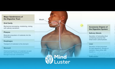
In human anatomy, the thigh is the area between the hip (pelvis) and the knee. Anatomically, it is part of the lower limb.
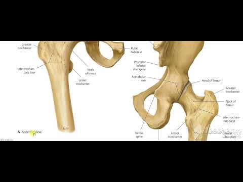
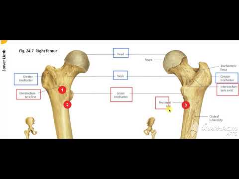
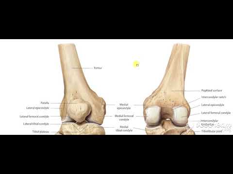
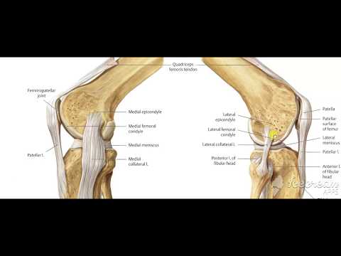
The single bone in the thigh is called the femur. This bone is very thick and strong (due to the high proportion of bone tissue), and forms a ball and socket joint at the hip, and a modified hinge joint at the knee
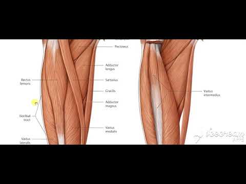
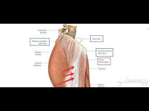
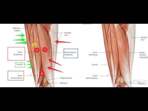
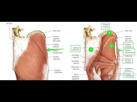
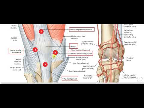
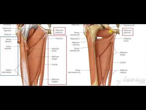
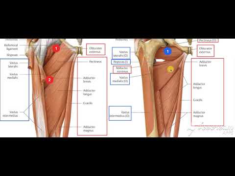
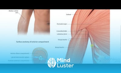
The musculature of the thigh can be split into three sections; anterior, medial and posterior. Each compartment has a distinct innervation and function.
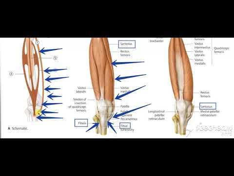
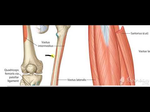
The muscles in the anterior compartment of the thigh are innervated by the femoral nerve (L2-L4), and as a general rule, act to extend the leg at the knee joint.
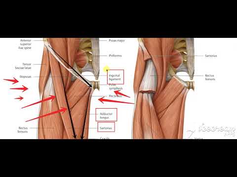
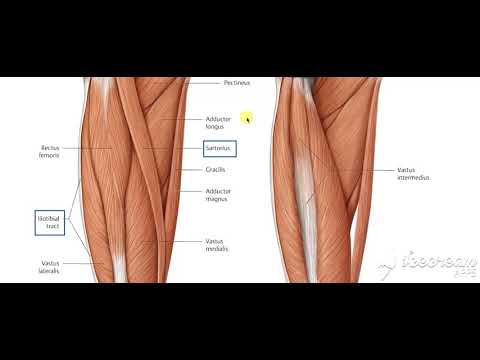
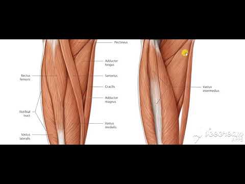
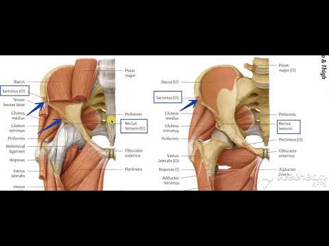
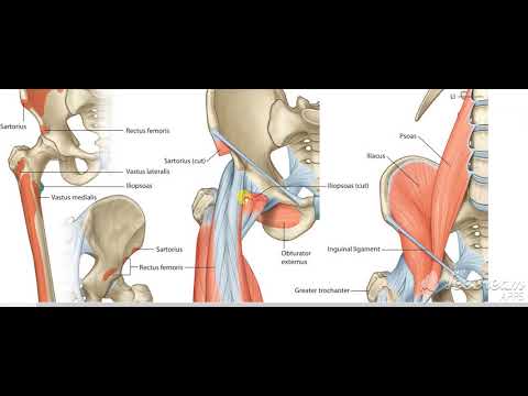
There are three major muscles in the anterior thigh – the pectineus, sartorius and quadriceps femoris. In addition to these, the end of the iliopsoas muscle passes into the anterior compartment.
This article will cover the attachments, actions, innervations and clinical correlations of these muscles.
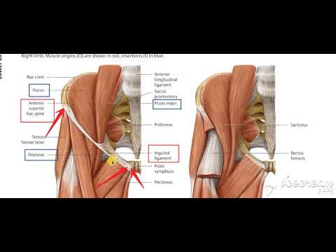
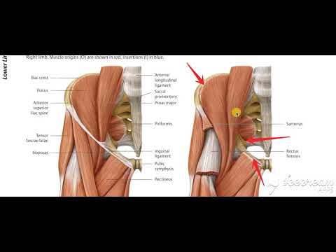
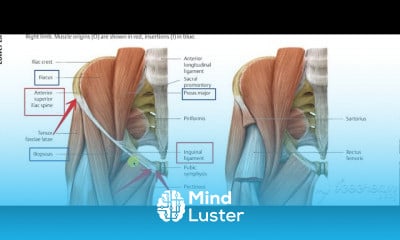
Iliopsoas
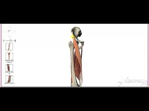
The iliopsoas is actually two muscles, the psoas major and the iliacus. They originate in different areas, but come together to form a tendon, hence why they are commonly referred to as one muscle.
Unlike many of the anterior thigh muscles, the iliopsoas does not extend the leg at the knee joint.
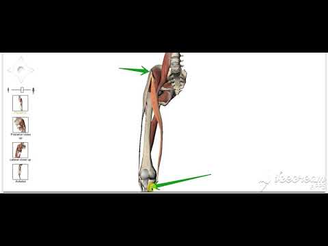
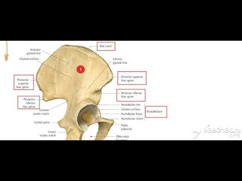
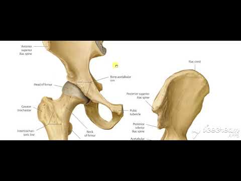
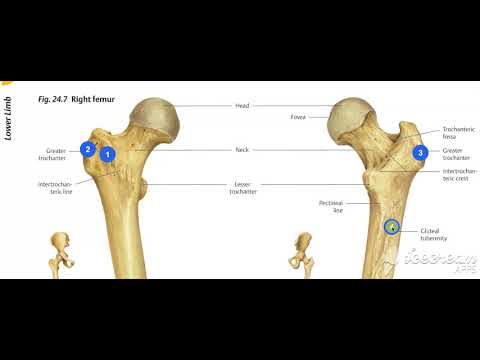
Attachments: The psoas major originates from the lumbar vertebrae, and the iliacus originates from the iliac fossa of the pelvis. They insert together onto the lesser trochanter of the femur.
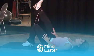
Actions: Flexes the thigh at the hip joint.
Innervation: The psoas major is innervated by anterior rami of L1-3, while the iliacus is innervated by the femoral nerve.