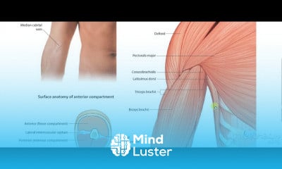
Vastus medialis muscle
Share your inquiries now with community members
Click Here
Sign up Now
Lessons List | 27
Lesson
Comments
Our New Certified Courses Will Reach You in Our Telegram Channel
Join Our Telegram Channels to Get Best Free Courses
Join Now
We Appreciate Your Feedback
2 Reviews
Vasco Sar

Harshini
Show More Reviews
Related Courses in Medical


Course Description
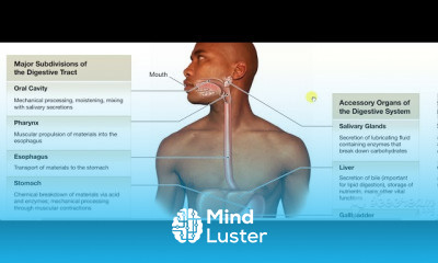
In human anatomy, the thigh is the area between the hip (pelvis) and the knee. Anatomically, it is part of the lower limb.
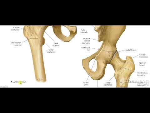
The single bone in the thigh is called the femur. This bone is very thick and strong (due to the high proportion of bone tissue), and forms a ball and socket joint at the hip, and a modified hinge joint at the knee
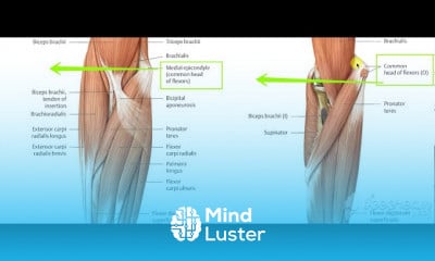
The musculature of the thigh can be split into three sections; anterior, medial and posterior. Each compartment has a distinct innervation and function.
The muscles in the anterior compartment of the thigh are innervated by the femoral nerve (L2-L4), and as a general rule, act to extend the leg at the knee joint.
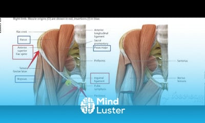
There are three major muscles in the anterior thigh – the pectineus, sartorius and quadriceps femoris. In addition to these, the end of the iliopsoas muscle passes into the anterior compartment.
This article will cover the attachments, actions, innervations and clinical correlations of these muscles.
Iliopsoas
The iliopsoas is actually two muscles, the psoas major and the iliacus. They originate in different areas, but come together to form a tendon, hence why they are commonly referred to as one muscle.
Unlike many of the anterior thigh muscles, the iliopsoas does not extend the leg at the knee joint.
Attachments: The psoas major originates from the lumbar vertebrae, and the iliacus originates from the iliac fossa of the pelvis. They insert together onto the lesser trochanter of the femur.
Actions: Flexes the thigh at the hip joint.
Innervation: The psoas major is innervated by anterior rami of L1-3, while the iliacus is innervated by the femoral nerve.
Trends
French
Data Science and Data Preparation
Formation efficace à l écoute de l
Graphic design tools for beginners
Artificial intelligence essentials
Learning English Speaking
Essential english phrasal verbs
MS Excel
Electrical engineering for engineer
Build a profitable trading
American english speaking practice
Build a tic tac Toe app in Xcode
YouTube channel setup
Design and Analysis of algorithms DAA
Python for beginners
Marketing basics for beginners
Figma for UX UI design
Magento Formation Français
Web Design for Beginners
Computer science careers
Recent
Data Science and Data Preparation
Growing ginger at home
Gardening basics
Ancient watering techniques
Grow mushrooms
Growing onions
Veggie growing
Bean growing at home
Growing radishes
Tomato growing at home
Shallot growing
Growing kale in plastic bottles
Recycling plastic barrel
Recycling plastic bottles
Grow portulaca grandiflora flower
Growing vegetables
Growing lemon tree
Eggplant eggplants at home
zucchini farming
watermelon farming in pallets