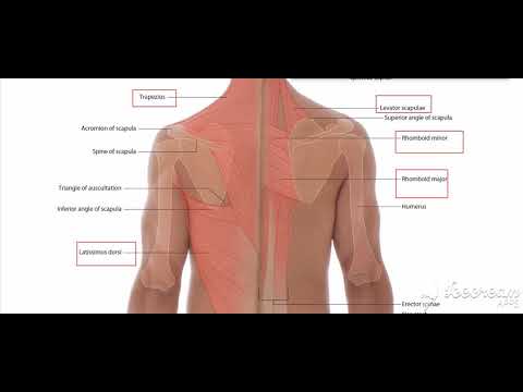
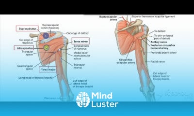
Trapezius muscle
Share your inquiries now with community members
Click Here
Sign up Now
Lessons List | 14
Lesson
Comments
Our New Certified Courses Will Reach You in Our Telegram Channel
Join Our Telegram Channels to Get Best Free Courses
Join Now
We Appreciate Your Feedback
30 Reviews

Rita Ndhlovu
Mirwais
Oddai Mohammad Abdullah abushash
Sarah Mokonyane
Show More Reviews
Related Courses in Medical

Course Description
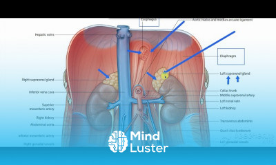
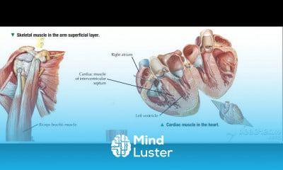
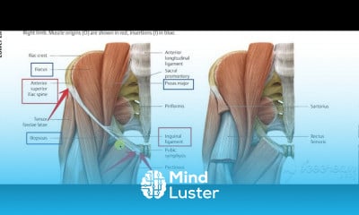
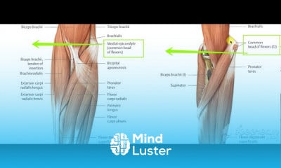
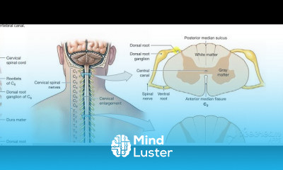
The relevant anatomy of the spinal nerve-muscular innervation of the back is centered around the lumbar spinal nerves, peripheral nerves of the lumbar plexus, spinal cord, and lumbar vertebral column. Within the lumbar region, the vertebral bodies are larger than in the thoracic and cervical regions due to the lumbar spine being designed for weight-bearing purposes. In general, the spinal cord consists of gray and white matter. As in the brain, the gray matter of the spinal cord contains the cell bodies; and the white matter of the spinal cord contains myelinated tracts. The gray matter of the spinal cord is found the central aspect of the spinal cord in the shape of the letter H. Immediately surrounding the spinal cord is the pia mater, with the subarachnoid space overlying the pia mater, the arachnoid mater overlying the subarachnoid space, and dura mater at the outermost layer, adherent to the spinal column.
Cerebral spinal fluid (CSF) is present in the central canal of the spinal cord in the center of the gray matter. Cerebral spinal fluid is also present surrounding the spinal cord, in the subarachnoid space and surrounding the spinal nerves. There are five lumbar vertebral bodies, five lumbar spinal nerves, and five lumbar spinal segments. The adult spinal cord terminates at the L1 or L2 vertebral level. The terminal aspect of the spinal cord is the conus medullaris, and immediately inferior to the spinal cord is the cauda equina. The cauda equina is a cordlike structure composed of thickened and elongated nerve roots that occupy the spinal canal. The cauda equina attaches to the mid-sacral canal at approximately the level of S2. Spinal nerves exit the spinal cord via the intervertebral foramen bilaterally at the lateral aspects of the vertebral column. Spinal nerves secure in place by thickenings in the pia mater, forming thin ligaments called denticulate ligaments. Denticulate ligaments attach to the arachnoid and dura mater stabilizing the position of each spinal nerve roots within the vertebral column
Trends
French
Graphic design tools for beginners
Printing student ID cards with excel tools
Artificial intelligence essentials
Essential english phrasal verbs
Build a profitable trading
MS Excel
Formation efficace à l écoute de l
Electrical engineering for engineer
Computer science careers
YouTube channel setup
Python programming language
Data Analytics Visualization Techniques
Back End Developer Learning Path course
Excel skills for math and science
English vocabulary with picture
Learning English Speaking
Python programming fundamentals A Z
Figma for UX UI design
Bioinformatics basics
Recent
Growing ginger at home
Gardening basics
Ancient watering techniques
Grow mushrooms
Growing onions
Veggie growing
Bean growing at home
Growing radishes
Tomato growing at home
Shallot growing
Growing kale in plastic bottles
Recycling plastic barrel
Recycling plastic bottles
Grow portulaca grandiflora flower
Growing vegetables
Growing lemon tree
Eggplant eggplants at home
zucchini farming
watermelon farming in pallets
pineapple farming