Middle ear site
Share your inquiries now with community members
Click Here
Sign up Now
Lessons List | 22
Lesson
Comments
Our New Certified Courses Will Reach You in Our Telegram Channel
Join Our Telegram Channels to Get Best Free Courses
Join Now
We Appreciate Your Feedback
8 Reviews

Omar shah
Nicodemus Isah
ABUBAKAR ABDULLAHI
Gezahegn legesse Shibeshi
Show More Reviews
Related Courses in Medical

Course Description
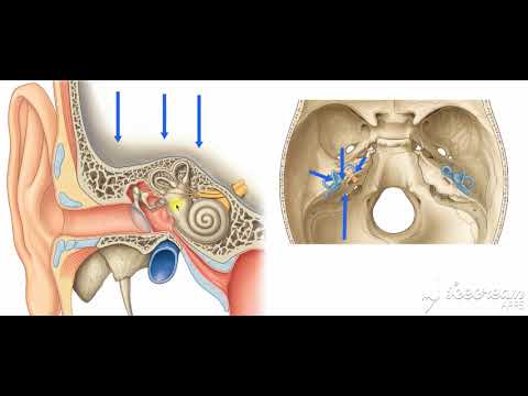
The ear is the organ that enables hearing and, in mammals, balance. In mammals, the ear is usually described as having three parts—the outer ear, the middle ear and the inner ear. The outer ear consists of the pinna and the ear canal. Since the outer ear is the only visible portion of the ear in most animals, the word "ear" often refers to the external part alone.The middle ear includes the tympanic cavity and the three ossicles. The inner ear sits in the bony labyrinth, and contains structures which are key to several senses: the semicircular canals, which enable balance and eye tracking when moving; the utricle and saccule, which enable balance when stationary; and the cochlea, which enables hearing. The ears of vertebrates are placed somewhat symmetrically on either side of the head, an arrangement that aids sound localisation.
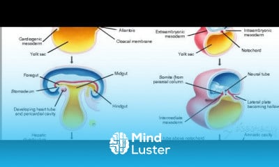
The ear develops from the first pharyngeal pouch and six small swellings that develop in the early embryo called otic placodes, which are derived from ectoderm.
The ear may be affected by disease, including infection and traumatic damage. Diseases of the ear may lead to hearing loss, tinnitus and balance disorders such as vertigo, although many of these conditions may also be affected by damage to the brain or neural pathways leading from the ear.
The ear has been adorned by earrings and other jewelry in numerous cultures for thousands of years, and has been subjected to surgical and cosmetic alterations.
The outer ear is the external portion of the ear and includes the fleshy visible pinna (also called the auricle), the ear canal, and the outer layer of the eardrum (also called the tympanic membrane).
The pinna consists of the curving outer rim called the helix, the inner curved rim called the antihelix, and opens into the ear canal. The tragus protrudes and partially obscures the ear canal, as does the facing antitragus. The hollow region in front of the ear canal is called the concha. The ear canal stretches for about 1 inch (2.5 cm). The first part of the canal is surrounded by cartilage, while the second part near the eardrum is surrounded by bone. This bony part is known as the auditory bulla and is formed by the tympanic part of the temporal bone. The skin surrounding the ear canal contains ceruminous and sebaceous glands that produce protective ear wax. The ear canal ends at the external surface of the eardrum.[3]
Two sets of muscles are associated with the outer ear: the intrinsic and extrinsic muscles. In some mammals, these muscles can adjust the direction of the pinna.In humans, these muscles have little or no effect. The ear muscles are supplied by the facial nerve, which also supplies sensation to the skin of the ear itself, as well as to the external ear cavity. The great auricular nerve, auricular nerve, auriculotemporal nerve, and lesser and greater occipital nerves of the cervical plexus all supply sensation to parts of the outer ear and the surrounding skin
The pinna consists of a single piece of elastic cartilage with a complicated relief on its inner surface and a fairly smooth configuration on its posterior surface. A tubercle, known as Darwin's tubercle, is sometimes present, lying in the descending part of the helix and corresponding to the ear-tip of mammals. The earlobe consists of areola and adipose tissue.The symmetrical arrangement of the two ears allows for the localisation of sound. The brain accomplishes this by comparing arrival-times and intensities from each ear, in circuits located in the superior olivary complex and the trapezoid bodies which are connected via pathways to both ears.
Trends
French
Graphic design tools for beginners
Artificial intelligence essentials
Formation efficace à l écoute de l
Essential english phrasal verbs
MS Excel
Build a profitable trading
Electrical engineering for engineer
YouTube channel setup
Python for beginners
Build a tic tac Toe app in Xcode
Magento Formation Français
Data Analytics Visualization Techniques
Web Design for Beginners
Excel skills for math and science
Learning English Speaking
Figma for UX UI design
Python programming language
Computer science careers
Marketing basics for beginners
Recent
Growing ginger at home
Gardening basics
Ancient watering techniques
Grow mushrooms
Growing onions
Veggie growing
Bean growing at home
Growing radishes
Tomato growing at home
Shallot growing
Growing kale in plastic bottles
Recycling plastic barrel
Recycling plastic bottles
Grow portulaca grandiflora flower
Growing vegetables
Growing lemon tree
Eggplant eggplants at home
zucchini farming
watermelon farming in pallets
pineapple farming