Posterior triangle floor
Share your inquiries now with community members
Click Here
Sign up Now
Lessons List | 36
Lesson
Comments
Our New Certified Courses Will Reach You in Our Telegram Channel
Join Our Telegram Channels to Get Best Free Courses
Join Now
We Appreciate Your Feedback
1 Reviews
Luis Martin Sussini
Show More Reviews
Related Courses in Medical

Course Description
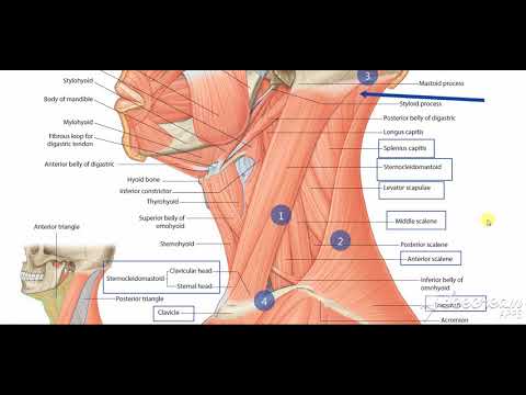
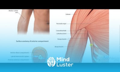
The anterior triangle is a region located at the front of the neck.
In this article, we shall look at the anatomy of the anterior triangle of the neck – its borders, contents and subdivisions.
Note: it is important to note that all triangles mentioned here are paired; they are located on both the left and the right sides of the neck.
Borders
The anterior triangle is situated at the front of the neck. It is bounded:
Superiorly – inferior border of the mandible (jawbone).
Laterally – anterior border of the sternocleidomastoid.
Medially – sagittal line down the midline of the neck.
Investing fascia covers the roof of the triangle, while visceral fascia covers the floor. It can be subdivided further into four triangles – which are detailed later on in this chapter.
The contents of the anterior triangle include muscles, nerves, arteries, veins and lymph nodes.
The muscles in this part of the neck are divided as to where they lie in relation to the hyoid bone. The suprahyoid muscles are located superiorly to the hyoid bone, and infrahyoids inferiorly.
There are several important vascular structures within the anterior triangle. The common carotid artery bifurcates within the triangle into the external and internal carotid branches. The internal jugular vein can also be found within this area – it is responsible for venous drainage of the head and neck.
Numerous cranial nerves are located in the anterior triangle. Some pass straight through, and others give rise to branches which innervate some of the other structures within the triangle. The cranial nerves in the anterior triangle are the facial [VII], glossopharyngeal [IX], vagus [X], accessory [XI], and hypoglossal [XII] nerves.
Suprahyoid Muscles Infrahyoid Muscles
Stylohyoid
Digastric
Mylohyoid
Geniohyoid
Omohyoid
Sternohyoid
Thyrohyoid
Sternothyroid
By TeachMeSeries Ltd (2021)
Fig 2 – The extracranial anatomical course of the hypoglossal nerve, through the anterior triangle of the neck.
Subdivisions
The anterior triangle is subdivided by the hyoid bone, suprahyoid and infrahyoid muscles into four triangles.
Carotid Triangle
The carotid triangle of the neck has the following boundaries:
Superior – posterior belly of the digastric muscle.
Lateral – medial border of the sternocleidomastoid muscle.
Inferior – superior belly of the omohyoid muscle.
The main contents of the carotid triangle are the common carotid artery (which bifurcates within the carotid triangle into the external and internal carotid arteries), the internal jugular vein, and the hypoglossal and vagus nerves.
By TeachMeSeries Ltd (2021)
Fig 3 – Carotid triangle of the neck
Clinical Relevance: Medical Uses of the Carotid Triangle
In the carotid triangle, many of the vessels and nerves are relatively superficial, and so can be accessed by surgery. The carotid arteries, internal jugular vein, vagus and hypoglossal nerves are frequent targets of this surgical approach.
The carotid triangle also contains the carotid sinus – a dilated portion of the common carotid and internal carotid arteries. It contains specific sensory cells, called baroreceptors. The baroreceptors detect stretch as a measure of blood pressure. The glossopharyngeal nerve feeds this information to the brain, and this is used to regulate blood pressure.
In some people, the baroreceptors are hypersensitive to stretch. In these patients, external pressure on the carotid sinus can cause slowing of the heart rate and a decrease in blood pressure. The brain becomes underperfused, and syncope results. In such patients, checking the pulse at the carotid triangle is not advised.
Submental Triangle
The submental triangle in the neck is situated underneath the chin. It contains the submental lymph nodes, which filter lymph draining from the floor of the mouth and parts of the tongue.
It is bounded:
Inferiorly – hyoid bone.
Medially – midline of the neck.
Laterally – anterior belly of the digastric
The floor of the submental triangle is formed by the mylohyoid muscle, which runs from the mandible to the hyoid bone.
By TeachMeSeries Ltd (2021)
Fig 4 – The submental triangle of the neck.
Submandibular Triangle
The submandibular triangle is located underneath the body of the mandible. It contains the submandibular gland (salivary), and lymph nodes. The facial artery and vein also pass through this area.
The boundaries of the submandibular triangle are:
Superiorly – body of the mandible.
Anteriorly – anterior belly of the digastric muscle.
Posteriorly – posterior belly of the digastric muscle.
By TeachMeSeries Ltd (2021)
Fig 5 – Lateral view of the neck, showing the submandibular triangle
Muscular Triangle
The muscular triangle is situated more inferiorly than the subdivisions. It is a slightly ‘dubious’ triangle, in reality having four boundaries. The muscular triangle contains some muscles and organs – the infrahyoid muscles, the pharynx, and the thyroid, parathyroid glands.
The boundaries of the muscular triangle are:
Superiorly – hyoid bone.
Medially – imaginary midline of the neck.
Supero-laterally – superior belly of the omohyoid muscle.
Infero-laterally – inferior portion of the sternocleidomastoid muscle.
By TeachMeSeries Ltd (2021)
Fig 6 – Muscular triangle of the neck.
Medicine on the Web
Ads By logo
img-responsive
Artificial Intelligence Predicts Survival 4 Ovarian Cancer
Medgoo.com
img-responsive
William Keimig, MD, Doesn’t Like to Think About Retirement
Medical Economics
img-responsive
We Need Help & Your Donation is 100% Tax-Deductible.
Docs without Borders
Quiz
The Anterior Triangle of the Neck
Question 1 of 3
Which of the following is NOT a border of the anterior triangle?
Anterior border of the sternocleidomastoid
Inferior border of the mandible
Sagittal line down the midline of the neck.
Anterior border of the sternohyoid
Report question
bookmarksRecommended reading
Aberrant Hypoglossal Nerve During Neck Dissection
Joshua D. Horton et al., JAMA Otolaryngology Head Neck Surgery
Contributions of Individual Muscles to the Submental Surface Electromyogram During Swallowing
Phyllis M. Palmer et al., Journal of Speech, Language, and Hearing Research, 1999
Cricopharyngeal Muscle Dysfunction Following Hypoglossal Nerve Stimulator Placement
Bryan J. Stevens et al., JAMA Otolaryngology Head Neck Surgery, 2018
New Jurassic mammaliaform sheds light on early evolution of mammal-like hyoid bones
Chang-Fu Zhou et al., Science, 2019
Powered by
person3D Model
Our 3D anatomical model provides you with hands-on, interactive and valuable learning tool right here on your device. To access the TeachMeAnatomy 3D Model, you must be a premium subscriber.
Trends
French
Graphic design tools for beginners
Data Science and Data Preparation
Formation efficace à l écoute de l
Artificial intelligence essentials
Learning English Speaking
Essential english phrasal verbs
MS Excel
Electrical engineering for engineer
American english speaking practice
Build a profitable trading
Build a tic tac Toe app in Xcode
Python for beginners
Figma for UX UI design
YouTube channel setup
Marketing basics for beginners
Magento Formation Français
Web Design for Beginners
Computer science careers
ArrayLists in C for beginners
Recent
Data Science and Data Preparation
Growing ginger at home
Gardening basics
Ancient watering techniques
Grow mushrooms
Growing onions
Veggie growing
Bean growing at home
Growing radishes
Tomato growing at home
Shallot growing
Growing kale in plastic bottles
Recycling plastic barrel
Recycling plastic bottles
Grow portulaca grandiflora flower
Growing vegetables
Growing lemon tree
Eggplant eggplants at home
zucchini farming
watermelon farming in pallets