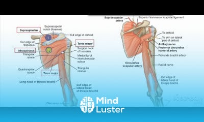
Back blood supply
Hide All Ads - Subscribe Premium Service Now
Share your inquiries now with community members
Click Here
Sign up Now
Lessons List | 14
Lesson
Comments
Our New Certified Courses Will Reach You in Our Telegram Channel
Join Our Telegram Channels to Get Best Free Courses
Join Now
We Appreciate Your Feedback
31 Reviews

AKSHAY GANGARAM GUPTA

Rita Ndhlovu
Mirwais
Oddai Mohammad Abdullah abushash
Show More Reviews
Related Courses in Medical

Course Description
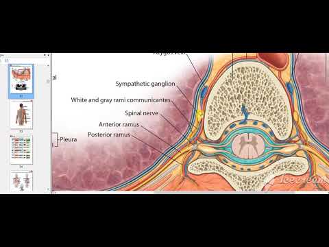
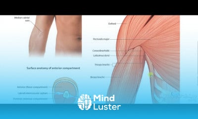
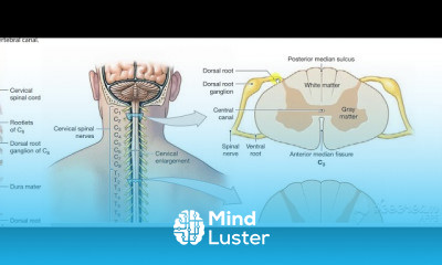
The relevant anatomy of the spinal nerve-muscular innervation of the back is centered around the lumbar spinal nerves, peripheral nerves of the lumbar plexus, spinal cord, and lumbar vertebral column. Within the lumbar region, the vertebral bodies are larger than in the thoracic and cervical regions due to the lumbar spine being designed for weight-bearing purposes. In general, the spinal cord consists of gray and white matter. As in the brain, the gray matter of the spinal cord contains the cell bodies; and the white matter of the spinal cord contains myelinated tracts. The gray matter of the spinal cord is found the central aspect of the spinal cord in the shape of the letter H. Immediately surrounding the spinal cord is the pia mater, with the subarachnoid space overlying the pia mater, the arachnoid mater overlying the subarachnoid space, and dura mater at the outermost layer, adherent to the spinal column.
Cerebral spinal fluid (CSF) is present in the central canal of the spinal cord in the center of the gray matter. Cerebral spinal fluid is also present surrounding the spinal cord, in the subarachnoid space and surrounding the spinal nerves. There are five lumbar vertebral bodies, five lumbar spinal nerves, and five lumbar spinal segments. The adult spinal cord terminates at the L1 or L2 vertebral level. The terminal aspect of the spinal cord is the conus medullaris, and immediately inferior to the spinal cord is the cauda equina. The cauda equina is a cordlike structure composed of thickened and elongated nerve roots that occupy the spinal canal. The cauda equina attaches to the mid-sacral canal at approximately the level of S2. Spinal nerves exit the spinal cord via the intervertebral foramen bilaterally at the lateral aspects of the vertebral column. Spinal nerves secure in place by thickenings in the pia mater, forming thin ligaments called denticulate ligaments. Denticulate ligaments attach to the arachnoid and dura mater stabilizing the position of each spinal nerve roots within the vertebral column
Trends
Graphic design tools for beginners
MS Excel
Learning English Speaking
Create cinematic ai landscapes videos
Python programming language
Google Python class
French
Create AI Videos
AWS For Beginners | Amazon AWS
Communication Skills
10X coding tools for developers
French language for beginners
Excel fundamentals for finance
Basic mathematics
American english speaking practice
Python machine learning from scratch
Workplace Communication skills for beginners
Logistic regression machine learning
Embedded Systems ES
Content marketing for beginners
Recent
RLC circuit basics
Types Of power diodes
HSPICE installation
Smart irrigation system
Python for data science fundamentals
Azure IoT webservices
Machine learning for beginners
IoT networking basics
SystemVerilog interview questions
Engineering chemistry fundamentals
Engineering thermodynamics
Mechanics
Electromagnetic theory
Thermometry
Basic mathematics
Create cinematic ai landscapes videos
Ai photography basics
Tools for ai image
Create ai videos with moonvalley
Leonardo Ai
You must have an account within the platform in order to participate in the discussion and comment. Register now for freeClick here