Course Description
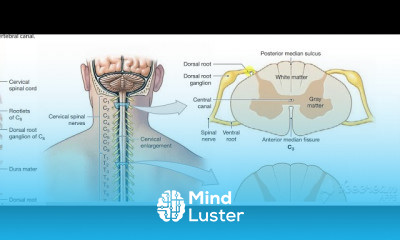
The relevant anatomy of the spinal nerve-muscular innervation of the back is centered around the lumbar spinal nerves, peripheral nerves of the lumbar plexus, spinal cord, and lumbar vertebral column. Within the lumbar region, the vertebral bodies are larger than in the thoracic and cervical regions due to the lumbar spine being designed for weight-bearing purposes. In general, the spinal cord consists of gray and white matter. As in the brain, the gray matter of the spinal cord contains the cell bodies; and the white matter of the spinal cord contains myelinated tracts. The gray matter of the spinal cord is found the central aspect of the spinal cord in the shape of the letter H. Immediately surrounding the spinal cord is the pia mater, with the subarachnoid space overlying the pia mater, the arachnoid mater overlying the subarachnoid space, and dura mater at the outermost layer, adherent to the spinal column.
Cerebral spinal fluid (CSF) is present in the central canal of the spinal cord in the center of the gray matter. Cerebral spinal fluid is also present surrounding the spinal cord, in the subarachnoid space and surrounding the spinal nerves. There are five lumbar vertebral bodies, five lumbar spinal nerves, and five lumbar spinal segments. The adult spinal cord terminates at the L1 or L2 vertebral level. The terminal aspect of the spinal cord is the conus medullaris, and immediately inferior to the spinal cord is the cauda equina. The cauda equina is a cordlike structure composed of thickened and elongated nerve roots that occupy the spinal canal. The cauda equina attaches to the mid-sacral canal at approximately the level of S2. Spinal nerves exit the spinal cord via the intervertebral foramen bilaterally at the lateral aspects of the vertebral column. Spinal nerves secure in place by thickenings in the pia mater, forming thin ligaments called denticulate ligaments. Denticulate ligaments attach to the arachnoid and dura mater stabilizing the position of each spinal nerve roots within the vertebral column