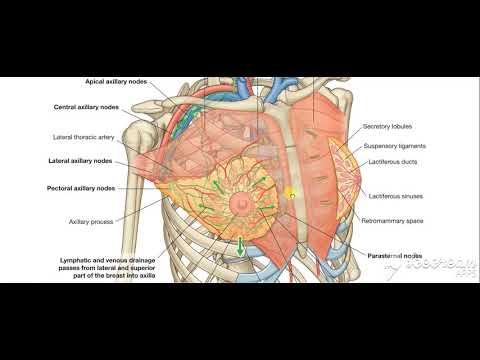
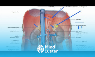
Breast blood supply Lymphatic drinage 2
Share your inquiries now with community members
Click Here
Sign up Now
Lessons List | 43
Lesson
Show More
Lessons
Comments
Our New Certified Courses Will Reach You in Our Telegram Channel
Join Our Telegram Channels to Get Best Free Courses
Join Now
We Appreciate Your Feedback
4 Reviews

Oussama Lanbache
Vasco Sar

Augusto Marcos Rodriguez Minella
Marim Omar dawood mohamed
Show More Reviews
Related Courses in Medical

Course Description
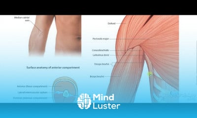
The pectoral region is the anterior region of the upper chest where there are four thoracoappendicular muscles (also known as the pectoral muscles):
pectoralis major
pectoralis minor
subclavius
serratus anterior
The breast is located superficial to the muscles. The lateral border of the pectoralis major muscle forms the anterior margin of the axilla. At the superolateral aspect the pectoral region is separated from the deltoid muscle by the deltopectoral groove and contains the cephalic vein.
What organs are in the pectoral region?
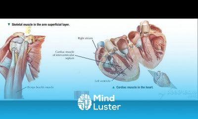
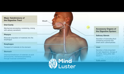
The chest is the area of origin for many of the body's systems as it houses organs such as the heart, esophagus, trachea, lungs, and thoracic diaphragm. The circulatory system does most of its work inside the chest.
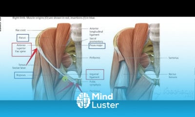
The pectoral region is located on the anterior chest wall. It contains four muscles that exert a force on the upper limb: the pectoralis major, pectoralis minor, serratus anterior and subclavius.
In this article, we shall look at the anatomy of the muscles of the pectoral region – their attachments, actions and innervation.
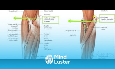
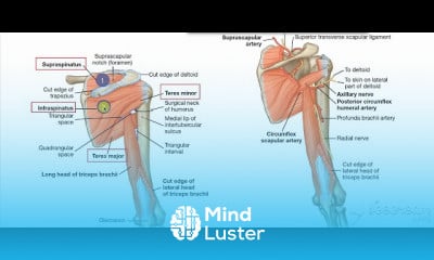
Pectoralis Major
The pectoralis major is the most superficial muscle in the pectoral region. It is large and fan shaped, and is composed of a sternal head and a clavicular head:
Attachments: The distal attachment of both heads is into the intertubercular sulcus of the humerus.
Clavicular head – originates from the anterior surface of the medial clavicle.
Sternocostal head – originates from the anterior surface of the sternum, the superior six costal cartilages and the aponeurosis of the external oblique muscle.
Function: Adducts and medially rotates the upper limb and draws the scapula anteroinferiorly. The clavicular head also acts individually to flex the upper limb.
Innervation: Lateral and medial pectoral nerves.
By TeachMeSeries Ltd (2021)
Fig 1 - The sternal and clavicular heads of the pectoralis major.
Fig 1 – The sternal and clavicular heads of the pectoralis major.
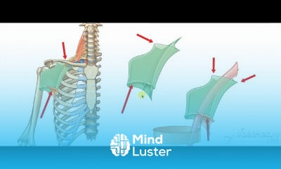
Pectoralis Minor
The pectoralis minor lies underneath its larger counterpart muscle, pectoralis major. Both muscles form part of the anterior wall of the axilla region.
Attachments: Originates from the 3rd-5th ribs and inserts into the coracoid process of the scapula.
Function: Stabilises the scapula by drawing it anteroinferiorly against the thoracic wall.
Innervation: Medial pectoral nerve.
Trends
French
Graphic design tools for beginners
Data Science and Data Preparation
Formation efficace à l écoute de l
Artificial intelligence essentials
Learning English Speaking
Essential english phrasal verbs
American english speaking practice
Electrical engineering for engineer
MS Excel
Build a profitable trading
Build a tic tac Toe app in Xcode
Python for beginners
Figma for UX UI design
YouTube channel setup
Magento Formation Français
Web Design for Beginners
Marketing basics for beginners
Computer science careers
ArrayLists in C for beginners
Recent
Data Science and Data Preparation
Growing ginger at home
Gardening basics
Ancient watering techniques
Grow mushrooms
Growing onions
Veggie growing
Bean growing at home
Growing radishes
Tomato growing at home
Shallot growing
Growing kale in plastic bottles
Recycling plastic barrel
Recycling plastic bottles
Grow portulaca grandiflora flower
Growing vegetables
Growing lemon tree
Eggplant eggplants at home
zucchini farming
watermelon farming in pallets