Parotid gland relations 4
Share your inquiries now with community members
Click Here
Sign up Now
Lessons List | 18
Lesson
Comments
Our New Certified Courses Will Reach You in Our Telegram Channel
Join Our Telegram Channels to Get Best Free Courses
Join Now
We Appreciate Your Feedback
10 Reviews

Bhagwat RAM Barar
Dr.Md.Abdur Rahman

Sohail Akram

Bimlendu Kumar Jha
Show More Reviews
Related Courses in Medical
Course Description
Parotid tumors are abnormal growths of cells (tumors) that form in the parotid glands. The parotid glands are two salivary glands that sit just in front of the ears on each side of the face. Salivary glands produce saliva to aid in chewing and digesting food.
There are many salivary glands in the lips, cheeks, mouth and throat. Tumors can occur in any of these glands, but the parotid glands are the most common location for salivary gland tumors. Most parotid tumors are noncancerous (benign), though some tumors can become cancerous.
Parotid tumors often cause swelling in the face or jaw that usually isn't painful. Other symptoms include numbness, burning or prickling sensations in the face, or a loss of facial movement.
Parotid tumor treatment is usually with surgery to remove the tumor. If the tumor contains cancer cells, additional treatments might be recommended.
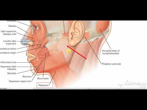
The parotid gland is a major salivary gland in many animals. In humans, the two parotid glands are present on either side of the mouth and in front of both ears. They are the largest of the salivary glands. Each parotid is wrapped around the mandibular ramus, and secretes serous saliva through the parotid duct into the mouth, to facilitate mastication and swallowing and to begin the digestion of starches. There are also two other types of salivary glands; they are submandibular and sublingual glands Sometimes accessory parotid glands are found close to the main parotid glands.
The parotid gland is a bilateral salivary gland located in the face. It produces serous saliva, a watery solution rich in enzymes. This is then secreted into the oral cavity, where it lubricates and aids in the breakdown of food.
In this article, we shall look at the location, vasculature and innervation of the parotid gland. We shall also consider any clinical correlations.
Anatomical Position
The parotid gland is a bilateral structure, which displays a lobular and irregular morphology. Anatomically, it can be divided into deep and superficial lobes, which are separated by the facial nerve.
It lies within a deep hollow, known as the parotid region. The parotid region is bounded as follows:
Superiorly – Zygomatic arch.
Inferiorly – Inferior border of the mandible.
Anteriorly – Masseter muscle.
Posteriorly – External ear and sternocleidomastoid.
The secretions of the parotid gland are transported to the oral cavity by the Stensen duct. It arises from the anterior surface of the gland, traversing the masseter muscle. The duct then pierces the buccinator, moving medially. It opens out into the oral cavity near the second upper molar.
Trends
Data Science and Data Preparation
Programming for Data Science with R
French
Artificial intelligence essentials
Electrical engineering for engineer
Build a profitable trading
Graphic design tools for beginners
American english speaking practice
Formation efficace à l écoute de l
Certified in CyberSecurity
Data Mining for Data Science
Learning English Speaking
Figma for UX UI design
Web Design for Beginners
Build a tic tac Toe app in Xcode
MS Excel
Essential english phrasal verbs
MIT Psychology
Marketing basics for beginners
Python for beginners
Recent
Qur anic reflections
Pillars of faith in islam
Pray in arabic word by word
Improve your Quran recitation
Read the Qur an
Islam in arabic basics
Qur an recitation
Arabic islamic words
Islamic vocabulary
Data Science and Data Preparation
Growing ginger at home
Gardening basics
Ancient watering techniques
Grow mushrooms
Growing onions
Veggie growing
Bean growing at home
Growing radishes
Tomato growing at home
Shallot growing